Haemophagocytic syndrome in a dog
History: A 4 year old female spayed Border Collie mix had a 10 day history of refractory IMHA/ ITP with possible DIC. She had abdominal effusion which was determined to be a modified transudate.
Gross Lesions:
Icterus of mucous membranes and mesentery. The abdominal cavity contained approximately 100-200 ml of bright orange clear fluid.
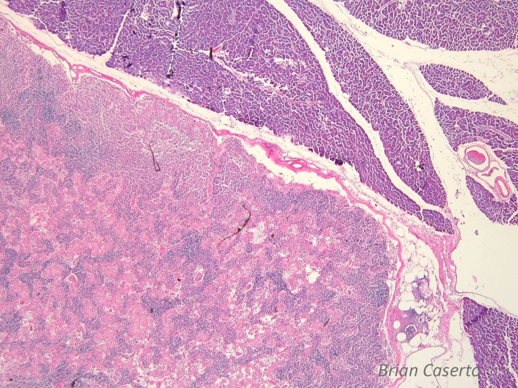
The spleen was diffusely enlarged with multifocal red to pink nodules.
The pancreas had a focal soft red nodule arising distal to the pyloric
junction.
Histopathology:
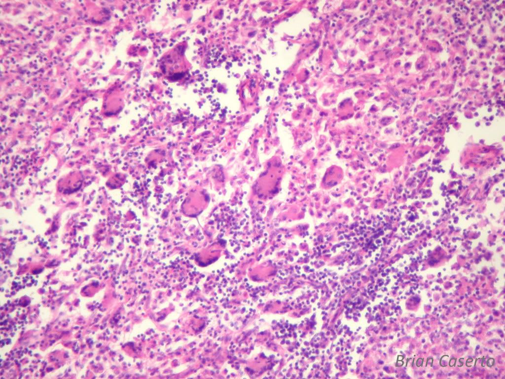
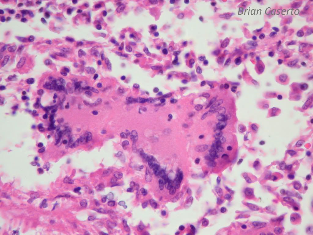
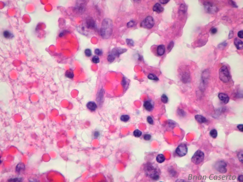
Lymph nodes: Normal lymph node architecture is replace and effaced by a mass of pleomorphic neoplastic histiocytes forming sheets of polygonal to spindle cells with ample eosinophilic cytoplasm and indented nuclei with vesicular chromatin. There are large clusters of multinucleated giant cells with up to 50-70 nuclei and large numbers of macrophages with intracytoplasmic erythrocytes (erythrophagocytosis), and large numbers of hemosiderophages. There is severe anisocytosis and anisokaryosis of the histiocytes and large numbers of bizarre nuclei.

Pancreatic lymph node: The lymph node is diffusely infiltrated by neoplastic histiocytic cells

Pancreatic lymph node: Numerous multinucleated neoplastic cells are present, with often bizarre shapes

Pancreatic lymph node: Multinucleated cells have nuclei too numerous to count, and often have bizarre mitotic figures

Pancreatic lymph node: There is prominent erythrophagocytosis by neoplastic histiocytic cells
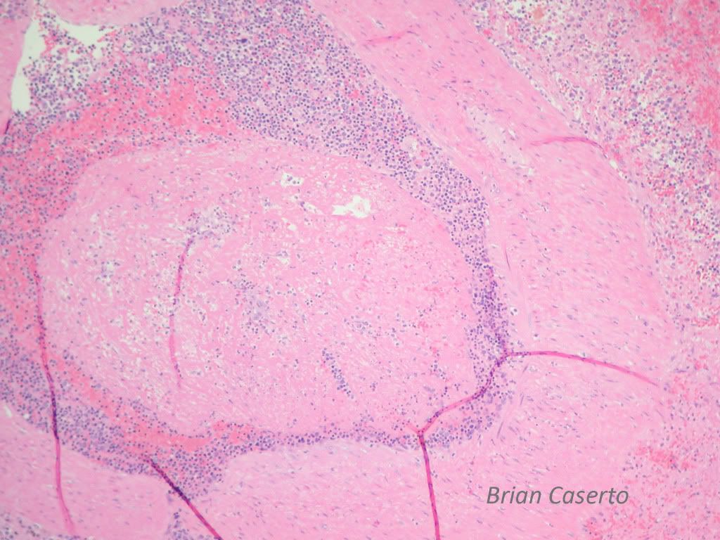
Spleen: There are multifocal areas of infarction and necrosis with intravascular fibrin thrombi in large arterioles. There is diffuse extramedullary hematopoiesis with large numbers of megakaryocytes and erythroid and myeloid precursors. Multifocal areas of the spleen are hypercellular.

Splenic artery: Occluding the lumen is a large fibrin thrombus
The liver and bone marrow contained small foci of neoplatic histiocytic cells.
Diagnosis:
1. Lymph nodes: Histiocytic sarcoma with erythrophagocytosis
2. Spleen: Multifocal infarction and marked extra-medullary
hematopoiesis
3. Bone Marrow: Multifocal histiocytic sarcoma, mild
4. Liver: Diffuse vacuolar degeneration with extra-medullary
hematopoiesis.
Comment:
The erythrophagocytosis seen histologically can be due either to the
IMHA or the histiocytic sarcoma or both. A hemophagocytic histiocytic
sarcoma has been described in dogs (Vet Pathol 43:632–645 (2006))
which is derived from splenic CD11d+ macrophages. This disease is
distinct from disseminated histiocytic sarcoma (malignant histiocytosis)
which is believed to arise from CD11c+ (CD11d-) dendritic cells and have
significantly less erythrophagocytosis as well as presenting with discrete
focal splenic nodules instead of having diffuse splenic enlargement. The
hemophagocytic syndrome mimics immune mediated hemolytic anemia
and thrombocytopenia but is Coombs negative. It presents with diffuse
splenic enlargement and can involve the liver and bone marrow and
lungs. But because they are similar in histological characteristics the best
way to differentiate the two diseases is with immunophenotyping on fresh
tissues.
About Brian
Anatomic Pathologist, VetPath Services, Stone Ridge, NY- musculoskeletal, oral/dental, and sinonasal diseases
www.vetpathservices.com