Canine Distemper Virus in 2 Racoons
History:
Racoon 1 was submitted for rabies testing, which was negative. No additional history is known.
Gross pathology: Unknown
Histopathology:
Racoon 1:
Brain: There is necrosis of numerous neurons, and intranuclear inclsions in neurons and astrocytes. Many blood vessels in the brain and the meninges are surrounded by moderate numbers of eosinophils with some lymphocytes and plasma cells.

Brain and meninges at low power. The meninges are expanded by eosinophils
Brain, high power: Numerous blood vessels are surrounded by moderate numbers of eosinophils

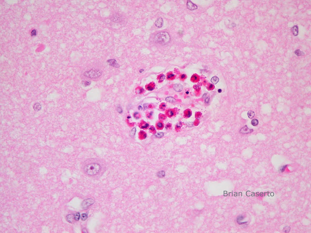
Brain: More perivascular eosinophils

Brain (labeled): Neuronal necrosis with nuclear inclusions in neurons and astrocytes
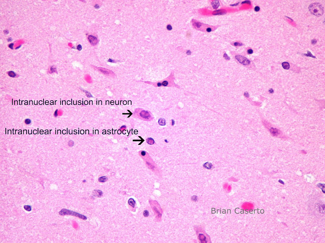
Brain with inclusions in the neurons and astrocyte nuclei
Spleen: Within the splenic capsule and muscular trabeculae and occasionally within the red and white pulp, there are large numbers of eosinophils.
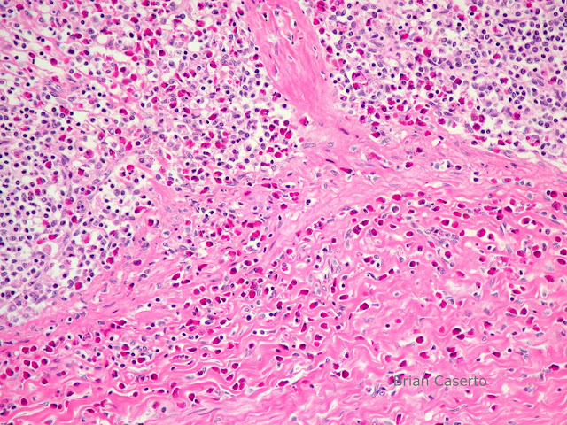
Spleen: Eosinophils in the capsule and in the stroma
Racoon 2:
Lungs: Bronchial and bronchiolar epithelium are multifocally necrotic and sloughing and large numbers contain eosinophilic intracytoplasmic inclusions. There are small numbers of epithelial viral syncytia among the respiratory epithelium. The lumen of bronchi and bronchioles contain sloughed epithelial cells and moderate numbers of neutrophils. The bronchiolar submucosa and peribronchial connective tissue contain moderate numbers of eosinophils.

Lungs, bronchioles: The epithelium is necrotic and sloughing with large numbers of intracytoplasmic inclusions
Lungs, bronchioles: There is a viral syncytial cell of bronchiolar epithelium
Lungs, bronchioles: Along with intracytoplasmic inclusions there are bronchioles with moderate numbers of degenerate and intact neutrophils
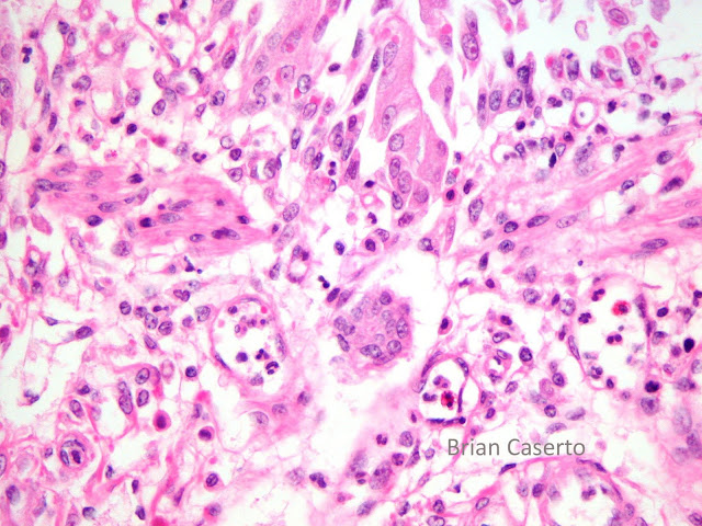
Renal pelvis: There are large numbers of intracytoplasmic eosinophilic inclusions in the urothelium, and at least one viral syncytia forming from transitional epithelial cells.

Renal pelvis: Degeneration of the transitional epithelium with cytoplasmic inclusions and a syncytial cell in the center
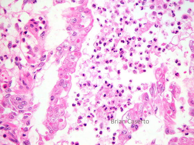
Stomach: The submucosa and lamina propria contain moderate numbers of eosinophils and few syncytia.
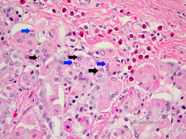
Stomach: Eosinophilic inclusions in the cytoplasm (BLUE) and nuclei (BLACK) of the mucosal epithelium
Liver: Diffusely the hepatocytes are swollen and the cytoplasm displaced by clear vacuoles of varying size.
There are no significant microscopic lesions in the brain, heart, kidneys, adrenal glands, lymph nodes, eyes, pancreas, or intestines.
Diagnosis:
Racoon 1:
Brain: Encephalitis and meningitis, eosinophilic with neuronal necrosis and intraneuronal and intraastrocytic inclusions
Spleen: Splenitis, eosinophilic, multifocal
Racoon 2:
1. Lungs: Necrotizing and suppurative bronchitis and bronchiolitis, moderate, multifocal, with intracytoplasmic epithelial eosinophilic inclusions and syncytia
2. Renal pelvis: Intracytoplasmic epithelial eosinophilic inclusions and viral syncytia
3. Stomach: Eosinophilic gastritis with intracytoplasmic epithelial eosinophilic inclusions
Comment:
Fluorescent antibody tests for CDV were positive in both cases. Racoon 1 was also positive by immunohistochemistry. The presence of eosinophils in both cases is intriguing since there was no histologic evidence of Toxoplasma or Baylisascaris infections which can be seen secondary to immunosuppression with canine distemper virus infections. The absence of zoites or sections of nematodes does not necessarily rule out infection by these agents.
One other case report of distemper in racoons found eosinophils in the lymph nodes and attributed them to the presence of large numbers of Baylisascaris procyonis in the intestines. The generalized presence of eosinophils with no obvious parasites or parasitic lesions in these cases may suggest an idiosyncratic inflammatory response to CDV. Lesions typically seen with Baylisascaris larva migration include linear tracts of malacia, gliosis, perivascular lymphoplasmacytic cuffing, and multifocal necrosis in other organs. Typical toxoplasma lesions include multifocal necrosis of numerous organs and in the brain, foci of necrosis and gliosis. Other conditions where eosinophils predominate include salt toxicity in pigs, some fungal infections, and occasionally Hairy Vetch toxicosis in bovines.
Reference:
M.R. CRANFIELD, I.K. BARKER, K.G. MEHREN AND W.A. RAPLEY. Canine Distemper in Wild Raccoons (Procyon lotor) at the Metropolitan Toronto Zoo. THE CANADIAN VETERINARY JOURNAL. Volume 25 (2) February 1984.