
Aplasia of the Major Duodenal Papilla or Bile Duct Atresia in a feline kitten
History: A 2 month old kitten died acutely with no previous clinical signs. FIV and FeLV tests were negative.
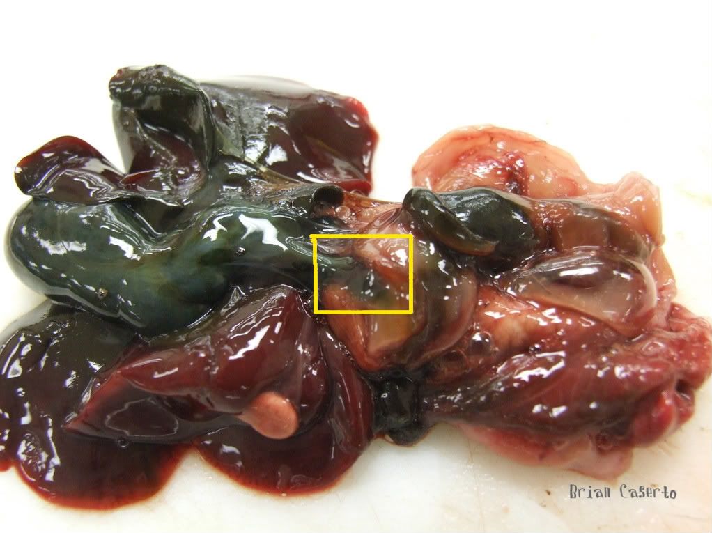
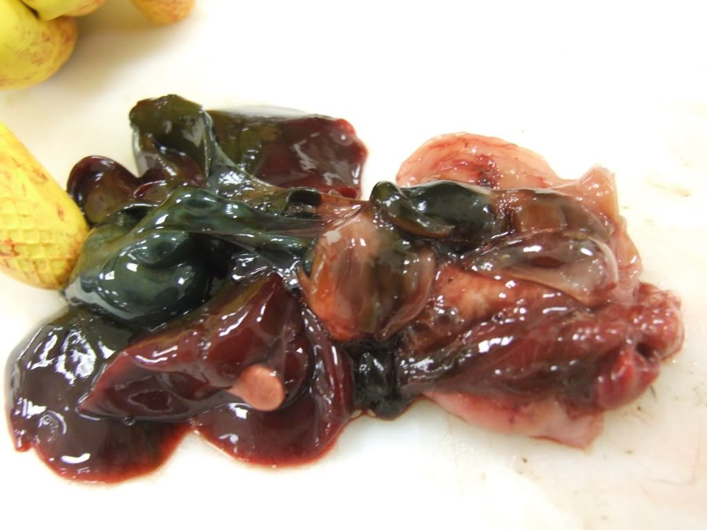
Gross Lesions: The kitten was very thin with little muscle mass or body fat stores. The gall bladder was markedly enlarged and dilated, as was the common bile duct all the way to the small intestine.
The bile duct was not patent and no duodenal papillae could be seen grossly. When the gall bladder was palpated the bile duct became more dilated at the serosal surface of the duodenum.
The bile duct ends blindly
In the photo above the bile duct is dilated and ends blindly at the serosa of the duodenum. In the picture below the gall bladder is being pressed and a small dilation can be seen at the duodenal serosa.

The Gall Bladder is pressed and a small dilation is visible at the duodenal serosa
In the photo below the duodenal lumen is shown which aside from the lack of a patent opening for the bile duct is otherwise normal.
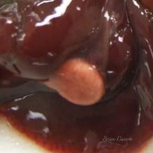
Mucosa of the duodenum. The bile duct (green) can be seen through the mucosa, but no opening is present
Histopathology:
The bile duct ends blindly in the tunica muscularis of the duodenum. The muscular layer is thinner where the bile duct enters, but is otherwise normal tissue. There is no sign previous injury to the tissue, or fibrosis. This leads me to believe that the opening did not form properly rather than becoming obstructed by disease.

Bile duct meeting the duodenum
In the picture above the white space in the upper left corner is the lumen of the bile duct. The white space in the lower right corner in the duodenal lumen. You can follow the muscular layer from left to right where it becomes thinner at the right edge of the photo. Just above the muscular wall is an area of necrosis and mild suppurative inflammation. There is no indication of fibrosis in the tissue.
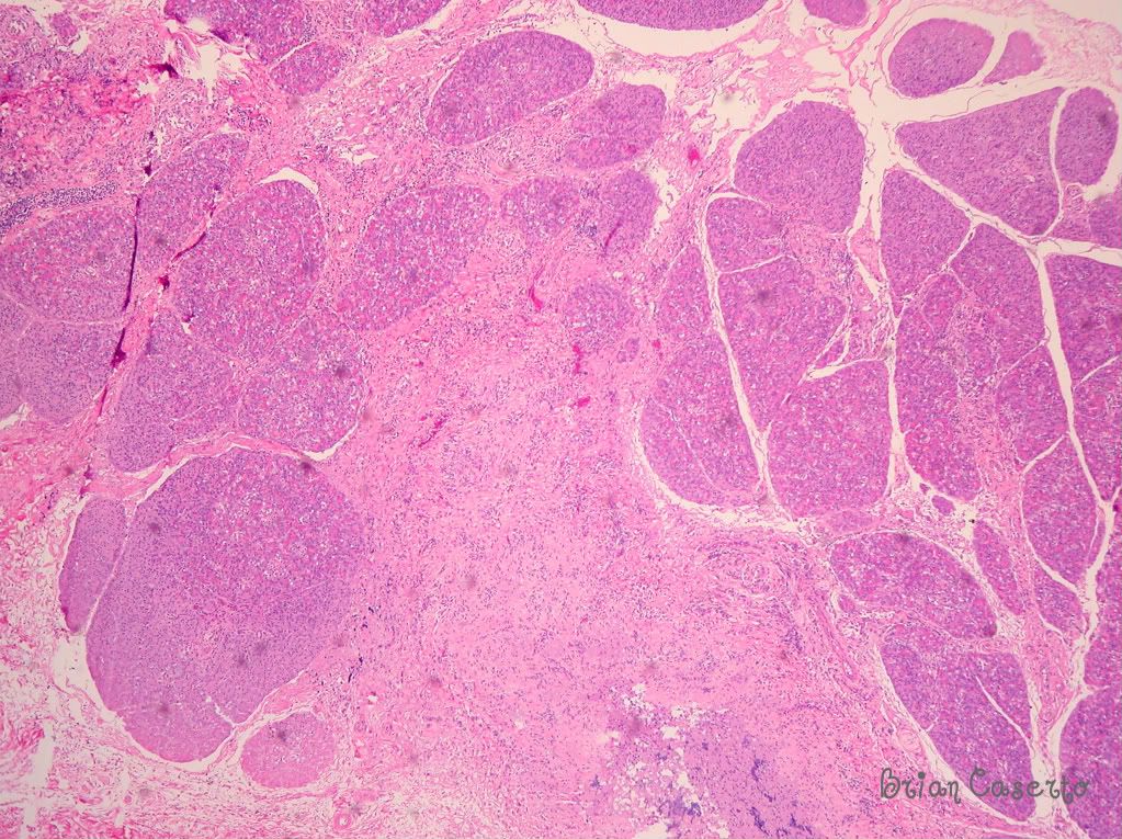
Pancreatic necrosis and inflammation
The pancreas (pictured above) had extensive areas of necrosis and suppurative inflammation around acini and in pancreatic ducts.
In cats the major duodenal papillae is the primary outlet for both Bile and Pancreatic secretions. Many cats do not even have a minor duodenal papillae like most dogs do. If the major papillae becomes blocked or is absent then both bile and pancreatic secretions will back up and cause tissue damage. In this case I think pancreatitis was secondary to obstruction of the outflow of pancreatic enzymes, and the necrosis seen in the above picture is due to both bile and pancreatic enzymes.
Incidental finding: The right adrenal gland was found not just adjacent to the liver, but joined to it via a small blood vessel arising from the liver.
Close up of the first image showing the adrenal gland attached to the liver


all the pictures are very nice & informative.
An alternative description of this condition could be Bile duct atresia/ Pancreatic duct atresia. This terminology acknowledges that the embyronic development of the hepatic diverticulum (which becomes the liver, gall bladder, and pancreas) had formed properly in the past. It may be that at some point in its maturation into the major duodenal papilla the diverticulum closes.
“The initial site of origin of the hepatic diverticulum is from the ventral side of the foregut. As the duodenum differentiates, unequal growth in the wall adjacent to where the hepatic diverticulum was located brings the origin of the diverticulum, now the entrance of the bile duct (thhe major duodenal papilla), to the dorsal surface of the duodenum…” (from Noden and deLahunta, Embryology of Domestic Animals, p 296-297, 1985, Williams and Wilkins)
Very nice and informative case! Great pictures!
I wonder if the cat could have had signs of rickets, as I am assuming he did not have properly absortion of Vit D…
Best regards!
Danilo Wasques
Nothing grossly evident, but I did not look at the bones histologically. A good thought, and If I come across anything similar I will post it.