Epidural steatitis and vertebral osteomyelitis in a dog
History: A 1 year old spayed female bulldog mix canine became acutely non-ambulatory, and both rear limbs were hyporeflexive.
Gross Necropsy:
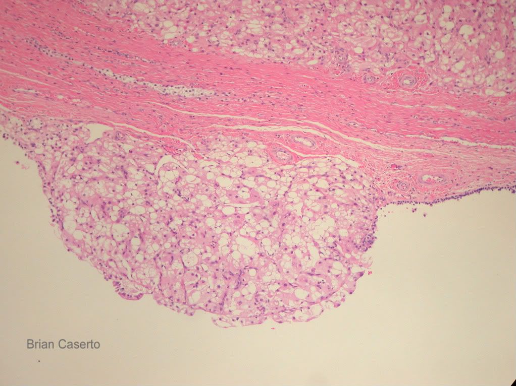
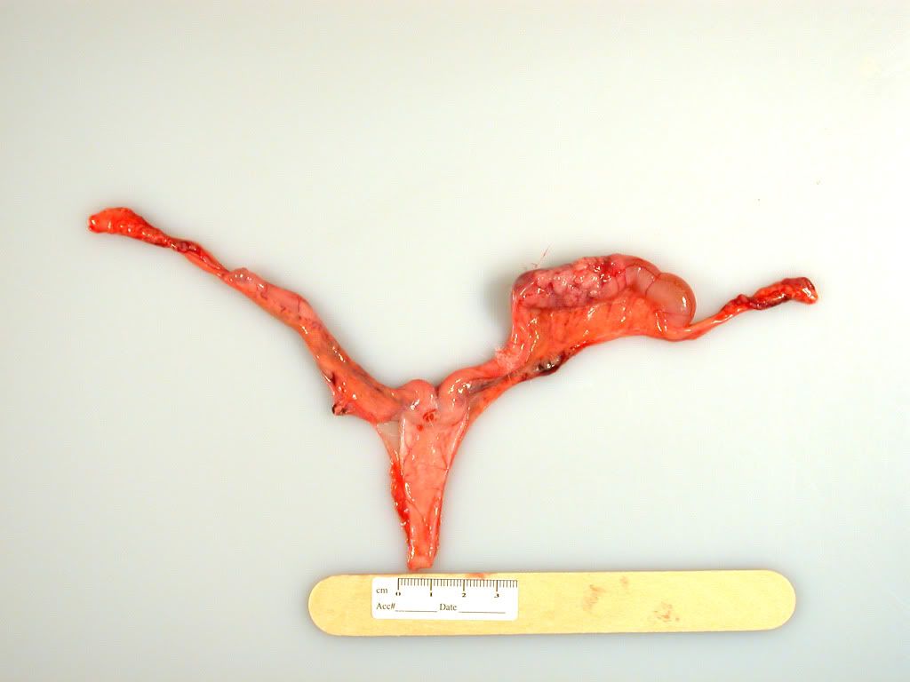
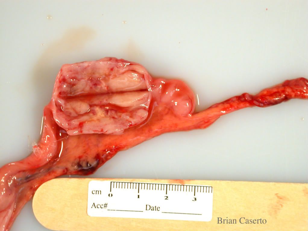
There was a 7 cm x 3 cm x 3 cm abscess in the epaxial musculature of the lumbar spine containing red-brown purulent fluid. The muscles surrounding the abscess including the epaxial and hypaxial musculature was pale, and there was a focal area of lysis of the ventral cortex of vertebral bodies L2 and L3.

Lumbar epaxial muscles: Abscess over spinal column
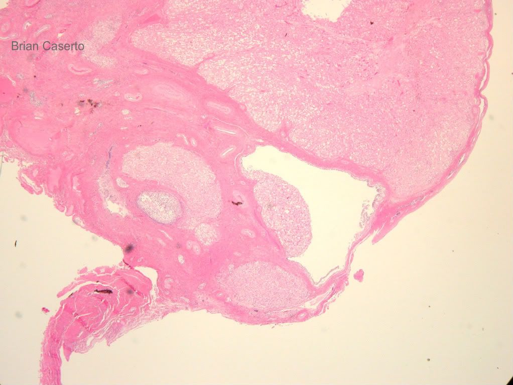
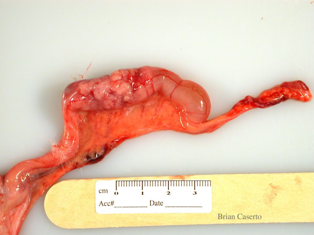
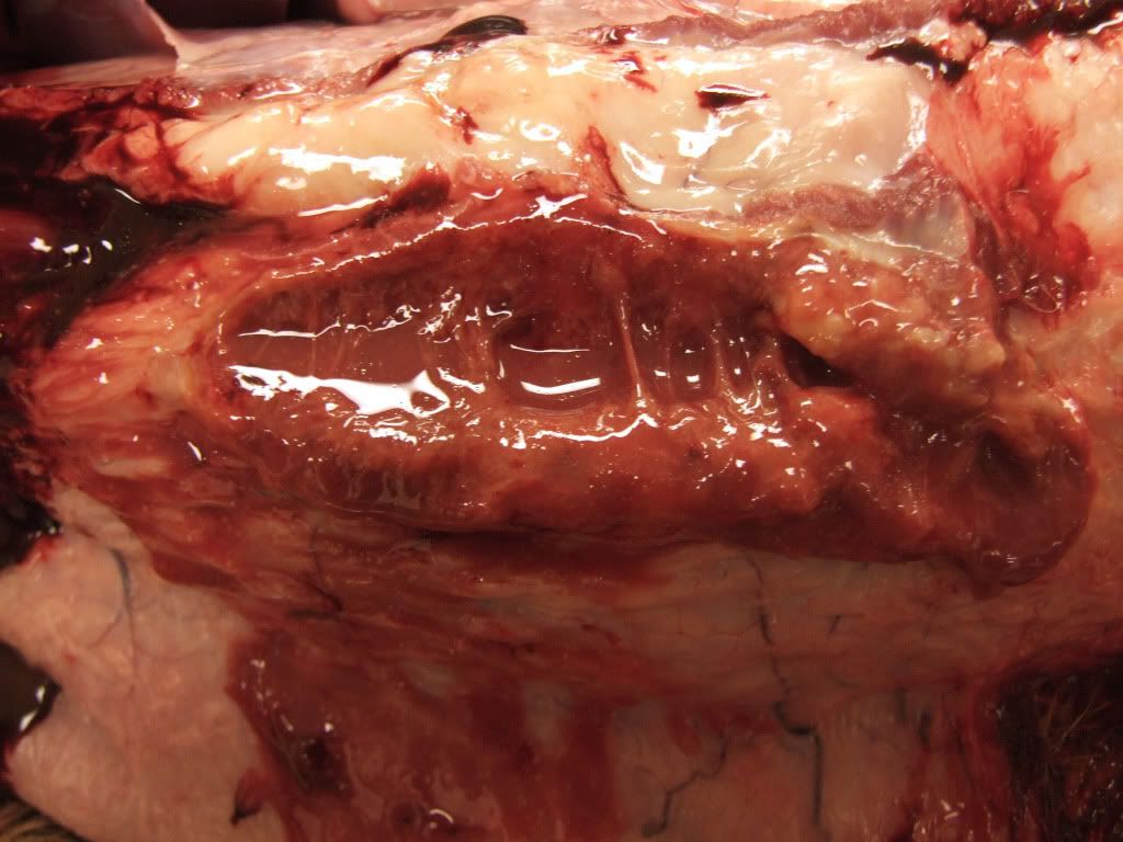
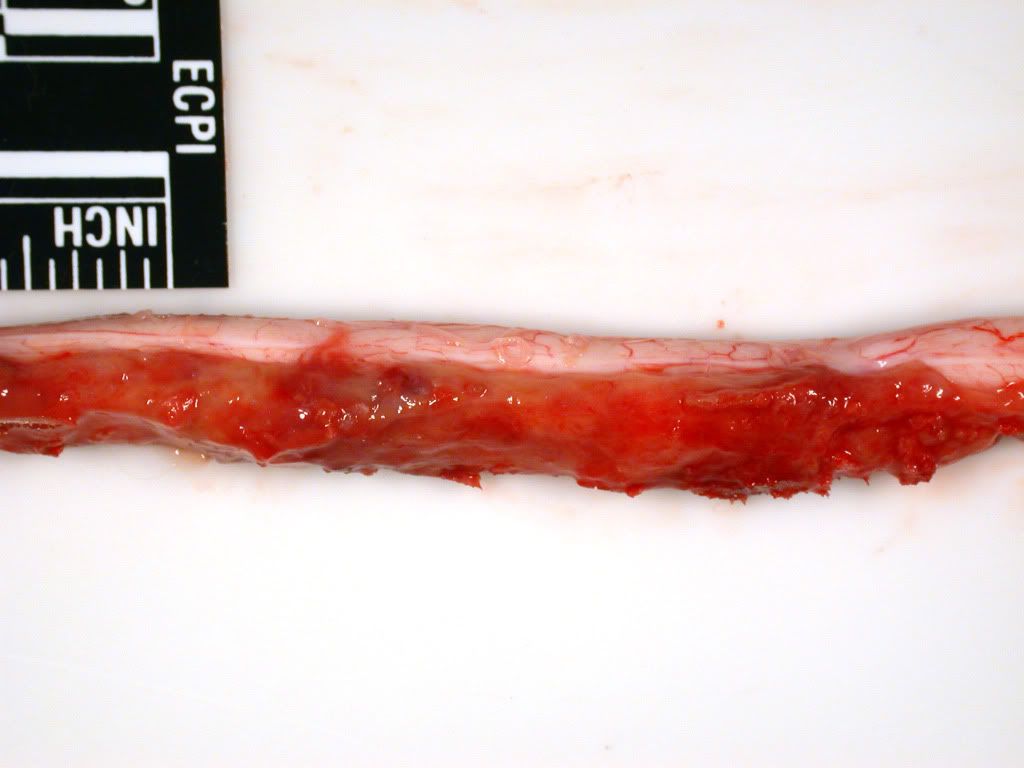
The dura of the spinal cord from vertebral bodies L2-L5 was covered with a thick layer of organized fibrin and suppurative material causing compression of the spinal cord. There were no gross lesions in the intervertebral discs and spinal cord.

Spinal cord: There is a thick mat of fibrinous and suppurative exudate lying on the spinal cord
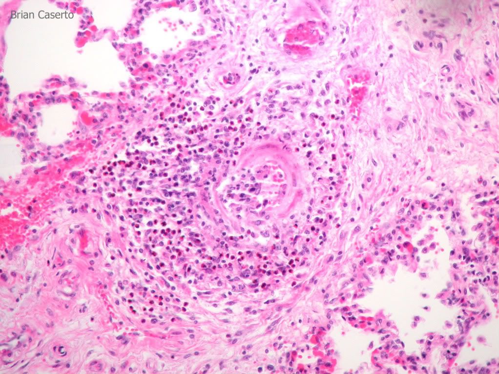
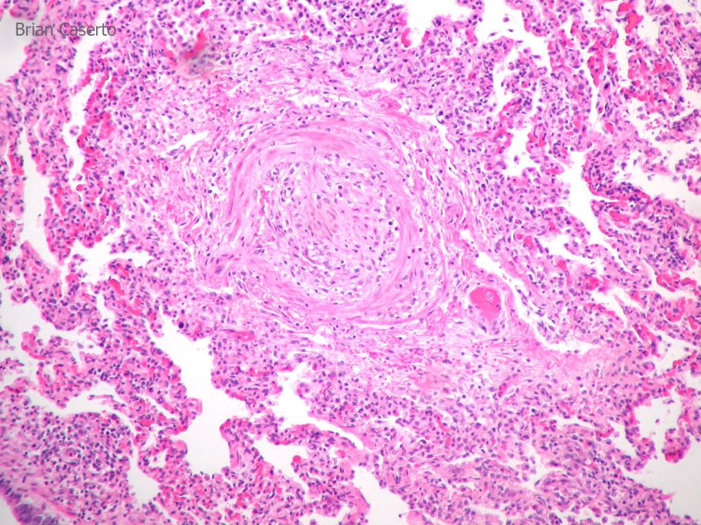
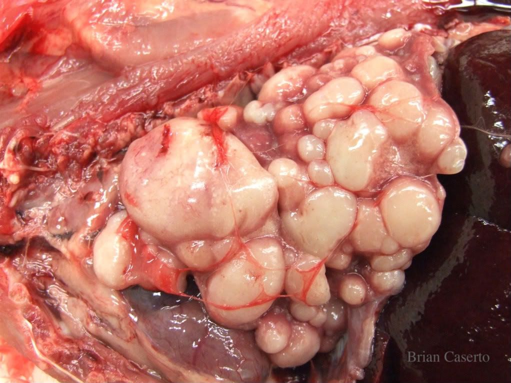
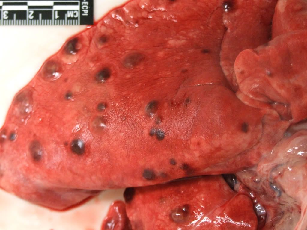
There were multifocal 3-5 mm round red nodules diffusely throughout the
lung.
There was a 7 cm x 3 cm x 3 cm abscess in the epaxial musculature of
the lumbar spine containing red-brown purulent fluid. The muscles
surrounding the abscess including the epaxial and hypaxial musculature
was pale, and there was a focal area of lysis of the ventral cortex of
vertebral bodies L2 and L3. The dura of the spinal cord from vertebral
bodies L2-L5 was covered with a thick later of organized fibrin and
suppurative material causing compression of the spinal cord. There were
no gross lesions in the intervertebral discs and spinal cord.
There were multifocal 3-5 mm round red nodules diffusely throughout the lung.

Lung: Multifocal embolic pneumonia
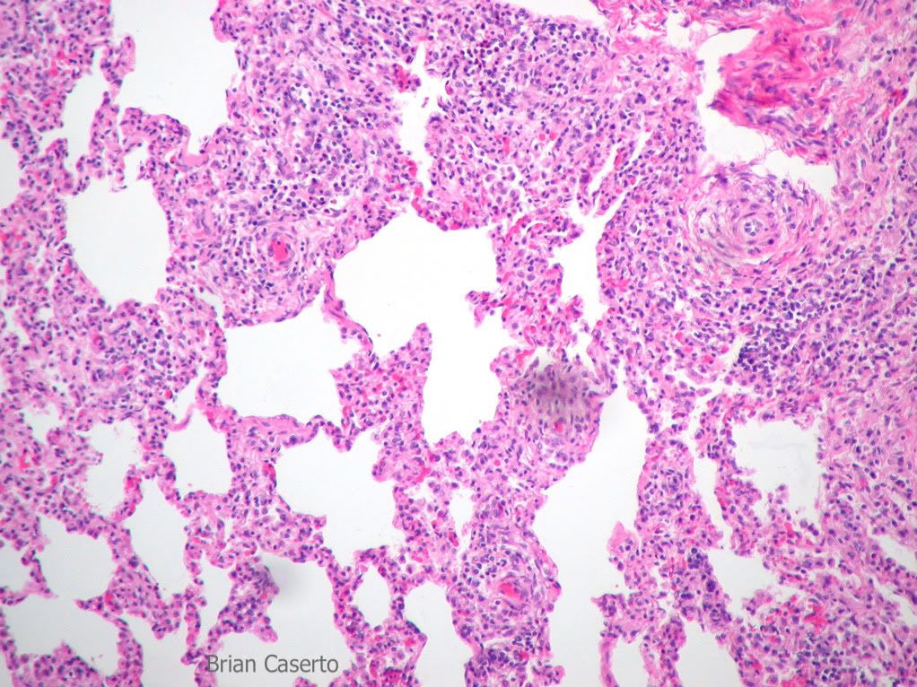
Histopathology:
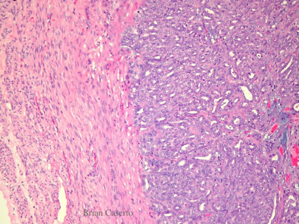
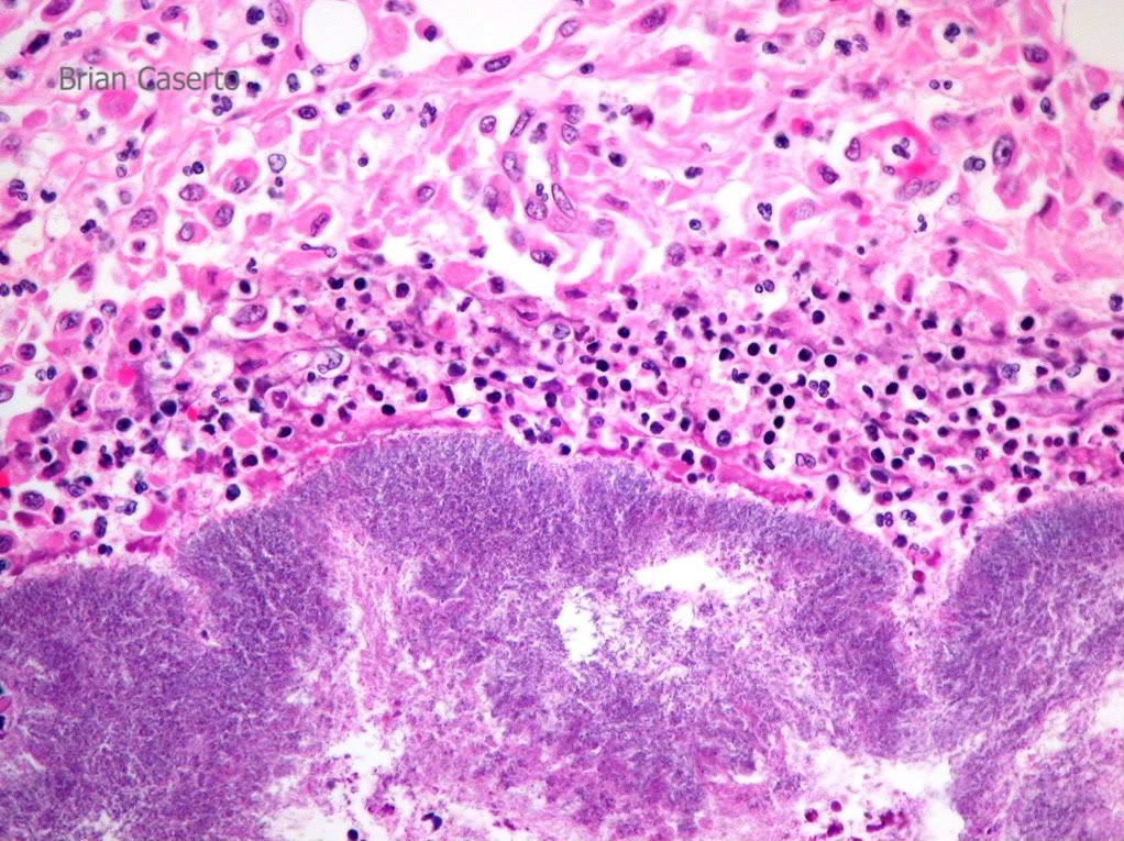
Epidural adipose: The epidural fat is infiltrated by large numbers of neutrophils, macrophages, lymphocytes and plasma cells with large amounts of fibrin and fibrous tissue. There are multiple bacterial colonies.

Spinal cord, dura and epidural adiopse- Suppurative steatitis
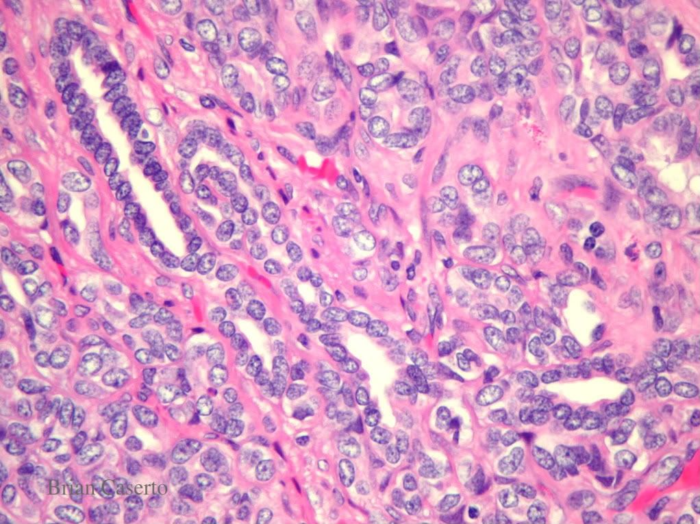
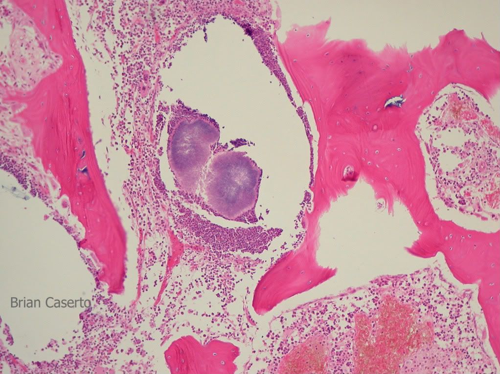
Vertebrae: The cortical bone is infiltrated and lysed by a large amount of neutrophils, macrophages and fibroblsats. There are large numbers of neutrophils surrounding bacterial colonies in the bone marrow of several vertebrae.

Vertebrae: Suppurative osteomyelitis with bacterial colonies
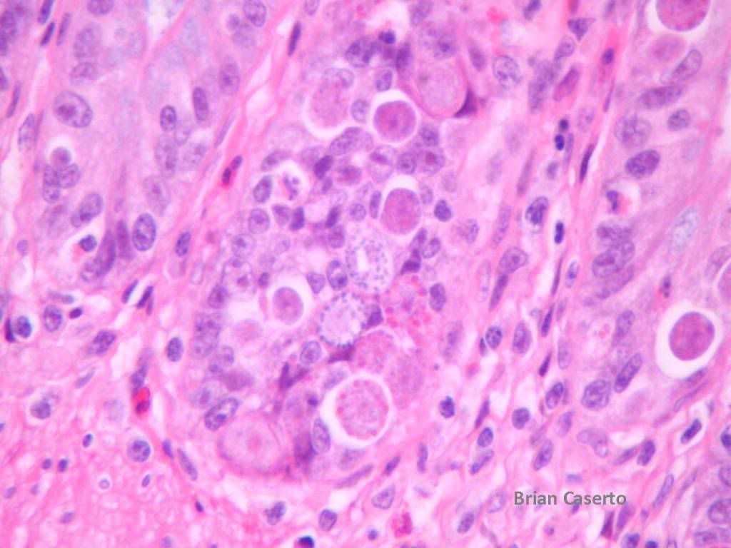
Bacterial colonies are composed of mixed gram positive rods and gram negative bacteria with the gram positive rods predominating. Similar bacterial colonies are present in the lungs and valvular thrombus.

Gram stain of bacteria- Large numbers of gram positive rods consistent with Campylobacter spp
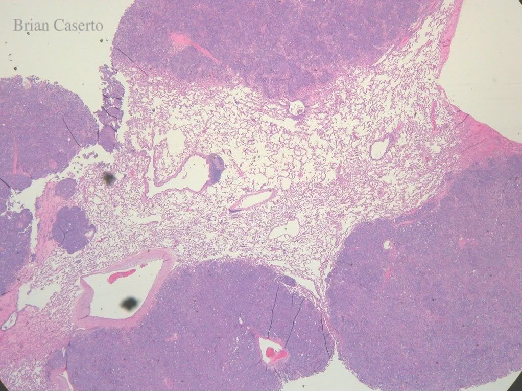
Aerobic culture: A swab from the exudate covering the spinal cord grew Corynebacterium spp and an unidentified bacteria.

Lung: Embolic suppurative pneumonia with bacterial colonies
Diagnosis:
1. Spinal Cord: Fibrinosuppurative epidural steatitis, chronic, focally extensive with compression of spinal cord
2. Vertebral body: Suppurative osteomyelitis with bacterial colonies
3. Lung: Multifocal suppurative pneumonia
4. Heart, right AV valve: Focal valvular endocarditis (not pictured)
Comment:
Microscopically the spinal column lesions are more chronic as evidenced by increased numbers of macrophages, lymphocytes, and fibroblast proliferation. Epidural steatitis can occur through direct extension of bacteria into the epaxial musculature or through hematogenous bacterial emboli. The infection spread into the vertebrae and epidural adipose tissue causing spinal cord compression and the neurologic signs noted in the history. Secondary bacteremia caused valvular endocarditis and embolic pneumonia.


You must be logged in to post a comment.